DXA is a low-cost, readily available imaging modality that is used globally to better understand Osteoporosis and other diseases and treatments
that impact bone health. With its
low cost and minimal radiation dose, DXA is ideally placed to enable researchers to
recruit large numbers of patients
and
perform multiple follow-up scans in a short time frame.
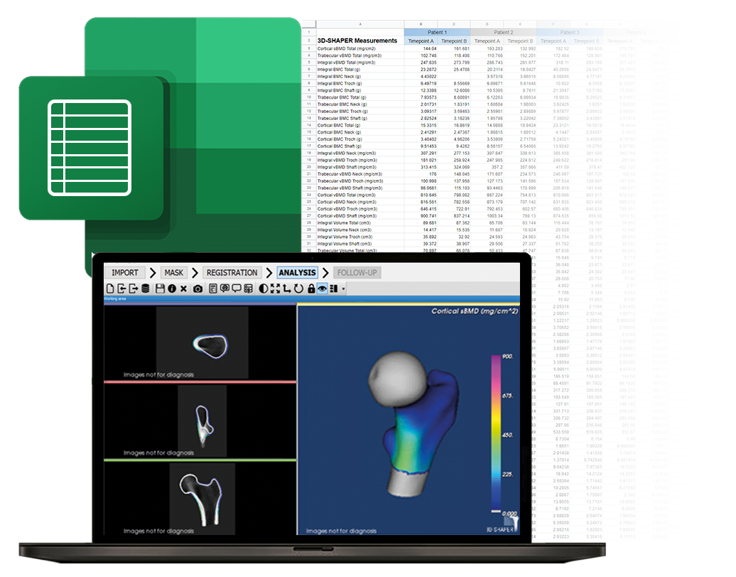
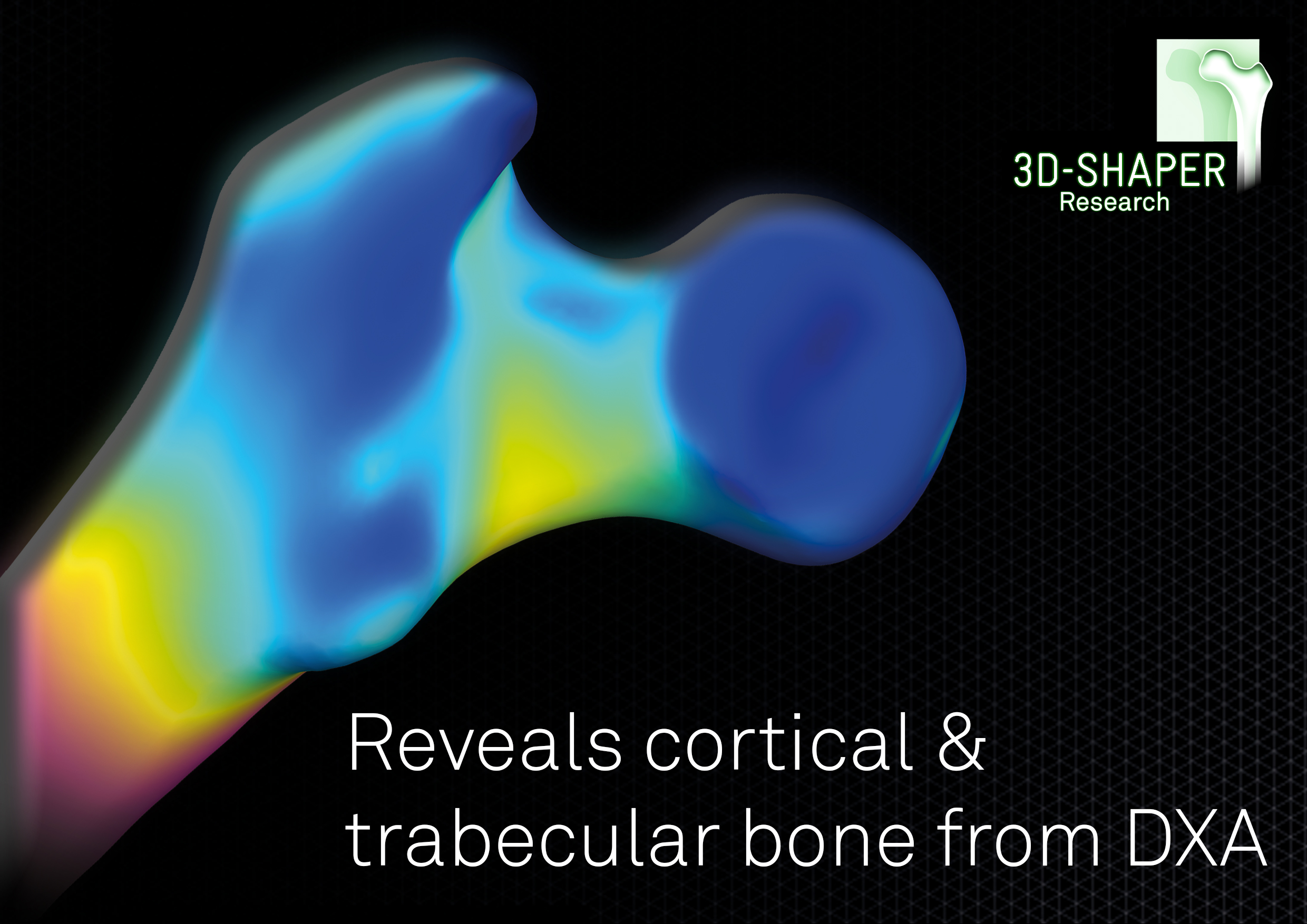
Despite its many advantages, DXA also brings limitations, as it only provides a two-dimensional assessment of bone density, known as areal BMD (aBMD).
This 2D representation of a 3D structure, whilst good,
limits the ability to delve deeper into the understanding of bone disease, lacking critical information on
the status and response of the cortical and trabecular bone compartments.
Maximize data insights while minimizing costs
But what if you could obtain QCT-like data from a routine DXA scan?
Well now you can with 3D-Shaper
® Research, the innovative software solution that bridges the gap between DXA and QCT-based assessments by providing
a patient-specific 3D QCT-like analysis from a standard 2D DXA image. That means
more patients, more time points and easier IRBs - all for a fraction of the cost.